After the 24th week of pregnancy, the fundal height matches your pregnancy week, plus or minus 3 centimeters, which is crucial for monitoring your baby’s growth. This measure, from your pubic bone to your uterus top, is key for checking your baby’s growth. Though it’s not perfectly precise, tracking the weeks pregnant your fundal in height helps doctors monitor how your baby develops.
Several risk factors though, like being overweight, having fibroids, or a full bladder, can tweak this measurement. These factors are critical in spotting issues like fetal growth restriction or twin pregnancies. If your fundal height is off, your doctor may request an ultrasound to investigate further.
Dr. Stan with Stork Advisor®️ has observed that while fundal height is helpful, it’s not always exact. This is why regular prenatal checks during pregnancy are very important. It is important to monitor growing trends to identify any discrepancies which may reflect differences with the baby’s normal development, either too much or too little.
Key Takeaways
- The fundal height generally matches the number of pregnancy weeks after 24 weeks, within a range of ±3 centimeters.
- Several factors—such as obesity and the presence of fibroids—can affect the accuracy of fundal height measurements.
- Changes in fundal height can signal important issues like oligohydramnios or polyhydramnios.
- Unusual fundal height measurements may necessitate an ultrasound for further investigation.
- Regular check-ups are vital for monitoring fetal growth and adjusting expectations as needed.
Introduction to Fundal Height and Fetal Size
Fundal height is an easy tool for tracking how much amniotic fluid and a baby grows during pregnancy. It measures the size of the uterus to check the amount of amniotic fluid and the baby’s growth. If the size seems off, it may hint at growth issues or if the baby is not in the right position. This makes health providers do more tests, like ultrasounds. Research suggests that 75% to 90% of fetal growth abnormalities go undiagnosed until birth.
One study compared measuring the fundal height to feeling the belly for baby’s size. The main findings, like the number of small-for-age babies or the death rate around birth, were similar. However, measuring the fundal height found small-for-age babies 30% to 50% of the time. Feeling the belly did it 48% of the time. This shows why it’s vital to monitor baby’s growth during pregnancy.
Fundal height tracking starts around 20 weeks into pregnancy. The measurement in centimeters should match the pregnancy length in weeks. This is a simple way to see if the baby is growing as expected.
Research shows that measuring fundal height can spot when there’s more than one baby, or if the baby is bigger than normal for its age. It can also show too much amniotic fluid or too little amniotic fluid. But, different people might measure it differently, which can make the results vary. Other things, like the mom’s body size and other health factors, can also affect the measurement. Ultrasounds are very good at spotting growth problems in the baby, with a success rate of about 93%.
- Measuring fundal height regularly helps catch any growth issues early on.
- Differences in measurements might happen due to things like the mom’s body size.
- Using both fundal height and ultrasound checks offers a clearer picture of the baby’s growth.
What is Fundal Height?
Fundal height measurement is a crucial part of monitoring a baby’s growth during pregnancy. Typically measured from around the 24th week of pregnancy, fundal height helps healthcare providers assess how well the baby is developing in the womb. This simple measurement offers valuable insights into the overall progress of the pregnancy.
Definition and Relevance
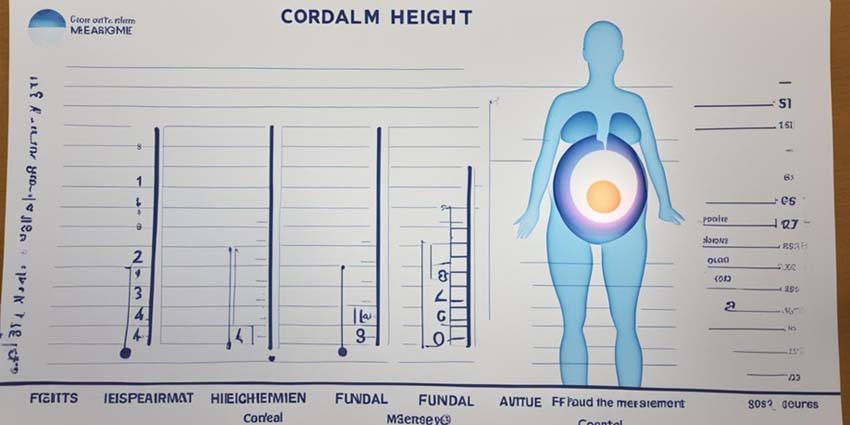
Fundal height is the distance from the top of the uterus (fundus) to the pubic bone. This measurement, taken in centimeters, usually corresponds to the number of weeks you are pregnant. For instance, at around 20 weeks of pregnancy, the fundal height typically reaches the level of the belly button. The accuracy of this measurement is vital for detecting potential issues in the pregnancy. Regular monitoring of fundal height can help identify growth problems in the baby, unusual levels of amniotic fluid, or the presence of multiple babies, such as twins. Pregnancy care providers typically start measuring fundal height routinely after the 24th week of pregnancy to ensure the baby is growing at a healthy rate.
Importance of Accurate Measurements
Accurate measurement of fundal height is essential for monitoring the health of both the mother and the baby. While small variations in size are generally considered normal, significant deviations from the expected measurements may require further investigation. For example:
- Excessive Fundal Height: If the fundal height is more than 3 centimeters larger than expected, it could indicate the presence of a large baby, uterine fibroids, or multiple pregnancies such as twins. This could also suggest an excessive amount of amniotic fluid (polyhydramnios), which may need to be monitored more closely.
- Reduced Fundal Height: On the other hand, if the fundal height is significantly less than expected, it could suggest potential growth restrictions in the baby, low levels of amniotic fluid (oligohydramnios), or issues with the placenta. These conditions could affect the baby’s development and may require medical intervention.
When discrepancies in fundal height measurements arise, healthcare providers often recommend further diagnostic tests, such as ultrasounds, to gain a more detailed understanding of the baby’s growth and the overall health of the pregnancy. Ultrasounds can provide more precise information on the baby’s size, amniotic fluid levels, and the condition of the placenta, allowing doctors to make informed decisions about the best course of action.
Conclusion
Fundal height measurement is a simple yet powerful tool in prenatal care, providing important insights into the baby’s growth and development. Regular and accurate measurement helps ensure that any potential issues are identified early, allowing for timely interventions that can support a healthy pregnancy. While slight variations in fundal height are common, significant deviations may signal the need for additional testing and closer monitoring. Understanding the importance of fundal height can help expectant mothers stay informed about their pregnancy progress and ensure the best possible outcomes for their baby.
How is Fundal Height Measured?
Fundal height is a crucial indicator used by healthcare providers to track your baby’s growth during pregnancy. It involves measuring the distance from the pubic bone to the top of the uterus (fundus). This measurement helps doctors estimate the baby’s size, monitor growth progress, and gauge how far along the pregnancy is, providing important information about the health and development of the baby.
The Measurement Process
The process of measuring fundal height typically begins around the 20th week of pregnancy and continues at every prenatal visit thereafter. During each visit, the healthcare provider will ask the mother to lie flat on her back. The doctor or midwife will then locate the pubic bone at the lower part of the abdomen and the top of the uterus (the fundus) at the upper part of the abdomen. Once these landmarks are identified, they will use a tape measure to determine the distance between these two points. This measurement, recorded in centimeters, is then compared to the number of weeks of pregnancy. Typically, the fundal height in centimeters should approximately equal the number of weeks of gestation, within a margin of plus or minus 2 centimeters.
For example, at 20 weeks of pregnancy, the fundal height should generally be around 18 to 22 centimeters. This measurement grows by about 1 centimeter per week as the pregnancy progresses. By 28 weeks, the fundal height usually measures around 28 centimeters, reflecting the steady growth of the baby. As the pregnancy nears full term, a slight drop in fundal height after 36 weeks might occur as the baby begins to descend into the pelvis in preparation for birth.
Factors That Affect Accuracy
While fundal height is a useful tool for monitoring fetal growth, several factors can influence the accuracy of the measurement. Variations in a mother’s body type, such as weight or height, can affect the measurement, as can the presence of uterine fibroids or the position of the baby within the uterus.
- Mother’s Weight and Height: A woman’s body composition can influence the measurement. For instance, a higher body mass index (BMI) might make it more challenging to palpate the top of the uterus, potentially leading to slight discrepancies in measurement.
- Uterine Fibroids: The presence of fibroids—noncancerous growths in the uterus—can alter the shape and size of the uterus, potentially leading to a higher or lower fundal height than expected.
- Baby’s Position: The baby’s position within the uterus can also cause variations in fundal height. For instance, if the baby is lying horizontally or if the baby has moved into the pelvis (especially in the third trimester), the measurement might be lower than expected.
- Amniotic Fluid Levels: Both excessive amniotic fluid (polyhydramnios) and low amniotic fluid (oligohydramnios) can affect fundal height measurements. Polyhydramnios might result in a higher measurement, while oligohydramnios could lead to a lower measurement.
Despite these potential variations, a difference of 1 to 2 centimeters from the expected fundal height is generally considered normal due to the natural changes in body positioning and baby movements.
Interpreting Fundal Height Measurements
Fundal height measurements provide valuable insight into fetal growth and development, but they are not infallible. A significantly lower-than-expected fundal height might indicate slow fetal growth, reduced amniotic fluid, or the baby moving into the pelvis earlier than anticipated. Conversely, a significantly higher fundal height could suggest multiple pregnancies (such as twins), an incorrect due date, or conditions like polyhydramnios.
Given these possible discrepancies, fundal height measurements are often used in conjunction with other diagnostic tools, such as ultrasounds. Ultrasounds can provide a clearer picture of the baby’s size, the amount of amniotic fluid, and the position of the placenta, all of which are critical for ensuring the well-being of both the mother and the baby.
Conclusion
Fundal height measurement is a simple yet important tool for monitoring the progress of a pregnancy. By regularly measuring the distance from the pubic bone to the top of the uterus, healthcare providers can track the baby’s growth and identify any potential concerns early on. While factors like maternal weight, uterine fibroids, and the baby’s position can influence the accuracy of the measurement, fundal height remains a valuable indicator when used in conjunction with other diagnostic methods. Regular and accurate measurement of fundal height helps ensure that the pregnancy is progressing as expected, allowing for timely interventions if needed to support the health and well-being of both mother and baby.
What Does Fundal Height Indicate About Your Baby?
Understanding fundal height is crucial during pregnancy, as it provides valuable insights into your baby’s growth and development. By regularly measuring fundal height, healthcare providers can track how well your baby is progressing each week, offering important clues about the baby’s health, size, and overall well-being.
Growth and Development
Fundal height is a reliable method used by doctors to gauge your baby’s growth during pregnancy. Typically, the measurement of fundal height begins around the 20th to 24th week of pregnancy. The amount pregnant your fundal height, measured in centimeters from the pubic bone to the top of the uterus (fundus), should approximately correspond to the number of weeks of pregnancy. For instance, at 28 weeks of gestation, the fundal height is generally around 28 centimeters.
This measurement is routinely monitored from around the 20th week until about the 36th week of pregnancy. During this period, the fundal height increases steadily, reflecting the baby’s continuous growth. By tracking this measurement, doctors can ensure that the baby is developing at a healthy rate. If the fundal height aligns closely with the gestational age, it generally indicates that the baby is growing well and on track developmentally.
Fundal height is particularly valuable because it provides an easy and non-invasive way to assess fetal growth. Regular monitoring of fundal height allows doctors to spot any deviations from the expected growth pattern early on. If the measurement doesn’t match up with the gestational age—for example, if it’s significantly higher or lower than expected—this could signal the need for additional tests, such as a prenatal ultrasound, to get a clearer picture of what’s happening inside the womb.
Potential Concerns
While fundal height is a useful tool for tracking pregnancy progress, it’s important to note that several factors can affect its accuracy. For example, the mother’s weight, height, and body type, as well as the baby’s position within the uterus, can influence the measurement. Despite these variations, fundal height remains a key indicator of how the baby is growing, and significant deviations from the expected measurement should be closely monitored.
- Lower Than Expected Fundal Height: If the fundal height is consistently lower than expected for the number of weeks pregnant, it might indicate that the baby isn’t growing as well as it should be. This could be due to various factors, such as intrauterine growth restriction (IUGR), which is when the baby is smaller than expected because of problems with the placenta, maternal health issues, or genetic conditions. A lower fundal height might also suggest low levels of amniotic fluid (oligohydramnios) or that the baby has moved into a position lower in the pelvis, especially as the pregnancy nears full term.
- Higher Than Expected Fundal Height: On the other hand, if the fundal height is significantly higher than the gestational age would suggest, this might indicate that the baby is growing larger than average, which could be a sign of gestational diabetes or other conditions. It could also suggest the presence of multiple babies (such as twins or triplets), or excessive amniotic fluid (polyhydramnios). In such cases, doctors may recommend additional prenatal ultrasounds to closely monitor the baby’s growth and assess the situation more accurately.
When any discrepancies measuring small or in fundal height are detected, further evaluation is typically required to ensure the health of both mother and baby. This might involve more frequent prenatal visits, ultrasounds, and possibly other diagnostic tests to determine the cause of the abnormal measurements. These steps are important for identifying and addressing any potential complications early, ensuring that both the mother and the baby receive the care they need.
Conclusion
Fundal height is a critical measure used during pregnancy to track your baby’s growth and ensure that they are developing as expected. Regular monitoring helps doctors assess whether the baby is growing at a healthy rate and can provide early warnings of potential issues. While variations in fundal height can occur due to factors like the mother’s body type or the baby’s position, significant deviations from the expected measurement may indicate underlying concerns that require further investigation. By keeping a close eye on fundal height, healthcare providers can ensure that any potential growth-related issues are addressed promptly, helping to safeguard the health and well-being of both mother and baby throughout the pregnancy.
Prenatal Ultrasound and Fundal Height
The journey of monitoring a pregnancy involves several tools and techniques, with fundal height measurements often serving as the initial step in assessing the baby’s growth. However, to gain a more comprehensive and accurate understanding of the baby’s development, prenatal ultrasounds play an essential role. While fundal height gives a general indication of the baby’s size and growth, ultrasounds provide a detailed and precise look at the baby’s physical development, position, and overall health. These insights are crucial for making informed health decisions throughout pregnancy.
The Role of Ultrasounds
Prenatal ultrasounds are indispensable in monitoring the health and development of the baby. These imaging tools allow healthcare providers to visualize the baby’s shape, check the amount of amniotic fluid surrounding the baby, and determine the location and health of the placenta. Unlike fundal height measurements, which provide a rough estimate of growth, ultrasounds offer a detailed, real-time image of the baby inside the womb.
Doctors rely on two main types of ultrasounds: transvaginal and transabdominal ultrasounds.
- Transvaginal Ultrasound: This type is typically used in the early stages of pregnancy, where the probe is inserted into the vagina to get a closer look at the developing fetus. It provides clearer images in the first trimester when the baby is still small.
- Transabdominal Ultrasound: This more common ultrasound method involves moving a probe across the abdomen to capture images of the baby. It’s frequently used throughout the pregnancy, especially in the second and third trimesters, to assess the baby’s growth, position, and overall health.
Studies have demonstrated that using a handheld ultrasound device to measure the baby’s abdominal circumference is particularly accurate, offering more reliable data than fundal height measurements alone. This precision underscores the importance of ultrasounds in prenatal care, as they allow for a detailed evaluation of the baby’s development that fundal height measurements alone cannot provide.
Ultrasounds are not just for assessing the baby’s growth but also for identifying any potential complications. They help monitor the amniotic fluid levels, detect any abnormalities in the baby’s development, and ensure the placenta is functioning properly. By providing such detailed information, ultrasounds guide crucial healthcare decisions during pregnancy, such as determining whether early delivery might be necessary or if additional interventions are required to support the baby’s health.
Frequency and Timing
The use of prenatal ultrasounds is carefully scheduled throughout the pregnancy to maximize their benefits while ensuring the safety of both the mother and baby. Typically, ultrasounds are performed at specific milestones in the pregnancy to gather important information:
- First Trimester Ultrasound (around 6-8 weeks): This early ultrasound confirms the pregnancy, determines the gestational age, and checks for the presence of more than one fetus. It also helps in identifying the baby’s heartbeat and assessing the overall health of the pregnancy.
- Second Trimester Ultrasound (around 18-22 weeks): Often referred to as the “anatomy scan,” this ultrasound is crucial for a detailed examination of the baby’s organs, bones, and overall structure. It’s also when the baby’s gender can often be determined if the parents wish to know.
- Third Trimester Ultrasound (around 28-40 weeks): Although not always routine, ultrasounds during this period may be conducted to monitor the baby’s position, the placenta’s health, and the amount of amniotic fluid. These ultrasounds are particularly important if there are concerns about the baby’s growth or if previous measurements, such as fundal height, indicate a potential issue.
During the third trimester, routine checks of fundal height might not always detect if a baby is smaller than expected for its gestational age, known as intrauterine growth restriction (IUGR). In such cases, doctors may recommend additional ultrasounds to obtain a clearer understanding of the baby’s condition. These extra ultrasounds help ensure that any growth concerns are identified and addressed promptly, allowing for timely interventions to support the baby’s health and development.
Conclusion
Fundal height measurements provide a useful starting point for assessing the baby’s growth during pregnancy, but prenatal ultrasounds are essential for gaining a full and accurate picture of the baby’s development. By combining the general growth trends indicated by fundal height with the detailed imagery provided by ultrasounds, healthcare providers can better monitor the baby’s health, identify potential issues, and make informed decisions to ensure a healthy pregnancy. The careful timing and frequency of ultrasounds throughout pregnancy allow for continuous monitoring and help to reassure both parents and the healthcare professional and providers that the baby is developing as it should. As a result, prenatal ultrasounds, alongside fundal height measurements, form a critical part of comprehensive prenatal care.
Symphysis-Fundal Height and Gestational Age
The Symphysis-Fundal Height (SFH) is key for figuring out how far along a pregnancy is. Sometimes, we’re not sure when the baby will arrive. By measuring from the pubic bone to the uterus top, doctors start this around week 24. This helps check if the baby grows right, based on the time in the womb.
SFH measurements follow the baby’s growth closely, going up steadily. Most women get checked five times during pregnancy. These checks are based on world standards to look for any growth issues. A study with over 13,000 women showed 92% had four or more checks. This helps in guessing the right age of the baby.
Measurements might vary a bit but getting them right is crucial. They spot problems like high blood pressure in pregnancy, sugar issues, or if there’s more than one baby. Research looked into how these measurements relate to baby health. They found no big difference in baby health issues compared to other methods.
The analysis made use of centile charts that adapt with the pregnancy. They kept it simple yet accurate. You can find the average and usual range of SFH at every pregnancy stage online at the INTERGROWTH-21st site. This helps doctors care for both mom and baby, avoiding health risks.
Monitoring Fetal Growth Through Pregnancy Measurements
Monitoring your baby’s growth is an essential component of a healthy pregnancy. Regular check-ups with your healthcare provider allow for close observation of your baby’s development, ensuring that any potential issues can be identified and addressed early. One of the primary methods for tracking your baby’s growth is Fetal Size Monitoring, which involves a combination of fundal height measurements and ultrasounds. These tools help doctors closely monitor your baby’s growth trajectory, allowing for timely interventions if necessary.
Regular Check-ups
Regular prenatal visits are crucial for monitoring your baby’s growth throughout pregnancy. During these visits, one of the most important measurements taken is the fundal height, which is the distance from the top of your uterus (fundus) to your pubic bone. This measurement, recorded in centimeters, typically corresponds to the number of weeks of pregnancy. For example, at 28 weeks of pregnancy, the fundal height should be around 28 centimeters, give or take a few centimeters. This simple yet effective measurement helps doctors assess whether your baby is growing as expected.
In addition to fundal height, ultrasounds are another key tool in monitoring fetal growth. These imaging tests provide a detailed view of the baby’s size, position, and overall development, giving healthcare providers more precise information than fundal height measurements alone. Regular monitoring through these methods is essential to detect any deviations from normal growth patterns.
Research conducted in countries like the UK, Australia, France, and the USA has shown that consistent monitoring of fetal growth is vital to ensure a healthy pregnancy outcome. Tracking your baby’s growth through these methods allows for early detection of potential problems, such as intrauterine growth restriction (IUGR) or macrosomia (when a baby is significantly larger than average), both of which can have serious implications for the pregnancy and delivery.
Understanding Fetal Growth Patterns
Understanding how a baby typically grows during pregnancy can help you appreciate the importance of regular monitoring. Fetal growth occurs in distinct phases:
- Up to 16 Weeks: In the early stages of pregnancy, a baby typically grows at a rate of about 19 grams per week. This steady but slower growth phase lays the foundation for the more rapid growth that occurs later.
- 16 to 20 Weeks: By the 20th week, the growth rate increases significantly to about 59 grams per week. This period marks a critical phase in the baby’s development, as organs and systems continue to mature.
- 20 to 30 Weeks: During this period, the baby’s growth rate accelerates further, reaching approximately 175 grams per week by the 30th week. This is when the baby’s size begins to significantly increase, with noticeable changes in the mother’s abdomen.
- 30 to 35 Weeks: Growth peaks during this time, with the baby gaining about 215 grams per week. This rapid growth is essential as the baby prepares for birth, with fat accumulation and continued organ development.
- 35 Weeks Onward: After the 35th week, the growth rate begins to slow slightly as the baby reaches the final stages of development. However, this is still a critical period for ensuring the baby is gaining enough weight and developing properly.
By monitoring these growth patterns, healthcare providers can assess whether your baby is following a healthy growth trajectory. If the growth appears to be slower or faster than expected, it may prompt additional testing to ensure that the baby is healthy and developing correctly.
Adjusting Growth Expectations
Throughout your pregnancy, your doctor will continually update their expectations for your baby’s growth based on the measurements taken at each visit. If the fundal height measurement deviates significantly from the expected range—specifically if it is off by more than 4 centimeters—this could indicate a potential issue that requires further investigation.
For example, a smaller-than-expected fundal height might suggest that the baby is not growing as quickly as it should, which could be due to factors such as placental insufficiency, low amniotic fluid, or intrauterine growth restriction (IUGR). Conversely, a larger-than-expected fundal height could indicate the presence of conditions such as gestational diabetes, polyhydramnios (excess amniotic fluid), or macrosomia (a large baby). In such cases, doctors may recommend additional ultrasounds or other diagnostic tests to gain a clearer understanding of the baby’s condition.
Ultrasounds are particularly useful in these situations, as they provide a more accurate assessment of the baby’s size and can help identify specific issues that might not be detected through fundal height measurements alone. While fundal height is a good general indicator of growth, ultrasounds can offer a detailed view, especially as the baby approaches full term. For instance, ultrasounds are often better at detecting smaller babies closer to birth, but they are also valuable for identifying larger babies or those in abnormal positions.
The timing and frequency of ultrasounds are carefully planned to ensure that doctors have the most accurate and up-to-date information about your baby’s growth without subjecting you to unnecessary tests. In the third trimester, when the baby’s growth is most rapid, more frequent ultrasounds may be recommended if there are any concerns about growth patterns. These additional checks help ensure that the baby is growing as expected and that any potential issues are addressed promptly.
Conclusion
Monitoring fetal growth through regular pregnancy measurements is essential for ensuring the health and well-being of both the mother and the baby. By combining fundal height measurements with the detailed imagery provided by ultrasounds, healthcare providers can closely track the baby’s development, detect any potential issues early, and make informed decisions to support a healthy pregnancy. Regular check-ups, an understanding of typical fetal growth patterns, and the ability to adjust growth expectations based on ongoing monitoring all play critical roles in creating the best conditions for your baby’s growth and health.
Addressing Concerns with Fetal Size
If the fundal height shows a fetus may be too small or large for its age, it’s important. Healthcare experts need to act quickly. Knowing about these issues helps manage possible complications.
Small for Gestational Age (SGA)
A fetus is tagged as Small for Gestational Age (SGA) if its birth weight is in the lowest 10% for its age. This often comes from Intrauterine Growth Restriction (IUGR), affecting up to 10% of all pregnancies. It’s a big cause of serious issues before and after birth.
Finding and handling IUGR is crucial. It helps avoid problems like stillbirth and long-term health issues. These can include troubles with brain development and heart diseases.
Large for Gestational Age (LGA)
A fetus is Large for Gestational Age (LGA) if its birth weight is in the highest 10%. A major issue with LGA is macrosomia, which leads to risks in delivery. These include shoulder dystocia and injuries during birth.
Macrosomia is a pregnancy complication often linked to the mother having gestational diabetes. Close watch and possible actions can handle these dangers well.
Studies Highlighting the Importance of Accurate Measurements:
- Pugh et al., 2018: They found a good way to guess the baby’s age at birth using fundal height and other body measures.
- Papageorghiou et al., 2016: They developed worldwide standards for measuring the height from the pubic bone to the top of the uterine fundus.
- Fung et al., 2020: They used computer algorithms to better predict how a baby grows.
Going to regular prenatal check-ups and addressing issues quickly is key for a safe pregnancy. Taking care of things like IUGR and macrosomia is vital during pregnancy.
Fundal Height and Fetal Size
The link between fundal height and baby size is key in prenatal care. Measuring from the pubic bone to the uterus top in centimeters estimates the fetus size. Doctors start this around week 20 and keep it up at every prenatal visit.
By week 24, fundal height should be close to how many weeks you are pregnant, give or take three centimeters. This regular check helps track the baby’s growth. Yet, being overweight or having fibroids could make these measures less accurate. After the 36th week, it’s harder to rely on fundal height, as it might not reflect true conditions like slow fetal growth or twins.
- Measuring more than three centimeters over could mean wrong due dates, obesity, or twins.
- Being more than three centimeters under might point to strong abs or growth issues inside the womb.
If measurements raise eyebrows, your doctor may suggest an ultrasound for a closer look at the baby’s size. While ultrasounds provide detailed images, they’re not always spot-on, typically off by 10 to 20 percent. Still, regular checks of fundal height give peace of mind about your baby’s growth and help catch any concerns early.
Conclusion
It’s vital for parents and health workers to grasp the importance of fundal height significance in antenatal growth assessment. It offers a quick peek at your baby’s growth. Though not perfect, it plays a big role in guiding more detailed checks for a healthy pregnancy.
Research indicates that single fundal height measurements might not always hit the mark, especially closer to delivery, missing the actual time by up to 4.4 weeks. Yet, taking 6-7 measurements can boost the accuracy to 95%. This method even works better than early ultrasound in some scenarios. Still, the method’s tendency to falsely signal slow growth (intrauterine growth restriction) cannot be ignored, highlighting the need for more tests.
Mixing fundal height checks with ultrasound readings increases their trustworthiness. A study highlighted by Rogers and Needham shows a tight link between fundal height and ultrasound, with significant correlation values. Even so, experts suggest starting these measurements at 25 weeks and doing them every 2-3 weeks, following health guidelines and global projects like “INTERGROWTH-21” that set international standards.
Regular check-ups using a mix of methods give the most precise picture of your baby’s growth. Understanding the role of fundal height in the context of prenatal growth checks is crucial. This knowledge helps protect the health of both the mother and the baby.
FAQ
What is fundal height and why is it important?
Fundal height is the distance from your pubic bone to the top of your uterus on top. It estimates the baby’s size and monitors growth. It also tracks the amniotic fluid after 24 weeks of pregnancy.
How is fundal height measured?
Healthcare providers find the pubic and pelvic bone, and uterus top. Then, they measure the space between with a tape. This happens at prenatal check-ups.
What are the factors affecting the accuracy of fundal height measurements?
Accuracy can be influenced by obesity, uterine fibroids, the baby’s position, and having more than one baby. These can alter the expected measurements.
What does an abnormal fundal height measurement indicate?
Abnormal measurements may suggest growth issues, too much amniotic fluid or too little amniotic fluid, or size concerns for the baby. An ultrasound might be needed to find out why.
How does fundal height correlate with gestational age?
After 24 weeks, the fundal height in centimeters should match the pregnancy weeks. For example, at 28 weeks, it’s around 28 centimeters.
Do you always need an ultrasound if the fundal height measurement is off?
Not always. If the measurement is unusual, an an ultrasound scan can help understand better. But it’s usually done only when necessary.
What is the role of prenatal ultrasound in relation to fundal height?
Ultrasounds give a detailed look at the baby’s size and growth. They check issues suggested by unusual fundal heights, like growth restrictions or being too large.
How often should fundal height be measured during pregnancy?
It’s measured at every prenatal visit after 24 weeks. Regular checks are key to watching the baby’s growth.
What is the significance of symphysis-fundal height in gestational age estimation?
It helps guess the baby’s age, especially when the due date is unclear. It’s a simple way to ensure the baby grows right, matching the timeline.
How do healthcare practitioners address concerns with fetal size based on fundal height measurements?
If measurements suggest the baby might be too small or big, more monitoring is done. Sometimes, actions are taken to handle any issues ensuring the baby’s well-being.
References
- What does fundal height measure in pregnancy? – https://www.mayoclinic.org/healthy-lifestyle/pregnancy-week-by-week/expert-answers/fundal-height/faq-20057962
- What to Know About Measuring Fundal Height – https://www.webmd.com/baby/what-to-know-about-measuring-fundal-height
- How Big Is My Baby? How to Measure Fundal Height and Fetus Size – https://www.whattoexpect.com/pregnancy/measuring-size-of-fetus
- Symphysial fundal height (SFH) measurement in pregnancy for detecting abnormal fetal growth – https://www.ncbi.nlm.nih.gov/pmc/articles/PMC6465049/
- Symphysis‐fundal height measurement in pregnancy – https://www.ncbi.nlm.nih.gov/pmc/articles/PMC7032650/
- What Is Fundal Height? – https://www.verywellhealth.com/fundal-height-7484340
- Fundal height measurement: What it means during pregnancy – https://www.babycenter.com/pregnancy/health-and-safety/fundal-height-measuring-large-or-small-for-gestational-age_1453305
- How to Measure Fundal Height – https://natalist.com/blogs/learn/how-to-measure-fundal-height?srsltid=AfmBOoqs0kamhGUwGfg4ntr-yteoT4yPwuQPa1xVqx5ku4ciKDugvKNi
- Fundal height: What it means, by week, and accuracy – https://www.medicalnewstoday.com/articles/fundal-height
- Twin Pregnancy and Fundal Height – OB-GYN New York City| Gynecologist, High Risk Pregnancy NYC – https://www.mfmnyc.com/blog/twin-pregnancy-and-fundal-height/
- Diagnostic accuracy of fundal height and handheld ultrasound-measured abdominal circumference to screen for fetal growth abnormalities – https://www.ncbi.nlm.nih.gov/pmc/articles/PMC4465094/
- Combination of Fundal Height and Ultrasound to Predict Small-for-Gestational Age at Birth – https://www.ncbi.nlm.nih.gov/pmc/articles/PMC8802337/
- International standards for symphysis-fundal height based on serial measurements from the Fetal Growth Longitudinal Study of the INTERGROWTH-21st Project: prospective cohort study in eight countries – https://www.bmj.com/content/355/bmj.i5662
- Symphysial fundal height (SFH) measurement in pregnancy for detecting abnormal fetal growth – Robert Peter, J – 2015 | Cochrane Library – https://www.cochranelibrary.com/cdsr/doi/10.1002/14651858.CD008136.pub3/full
- What Is Symphysis Fundal Height (SFH)? – https://www.sommersandroth.com/blog/uncategorized/what-is-symphysis-fundal-height-sfh/
- Monitoring fetal growth – NCBI Bookshelf – https://www.ncbi.nlm.nih.gov/books/NBK573939/
- Fetus size by week: Your baby’s weight throughout pregnancy – https://www.babycenter.com/pregnancy/your-body/growth-chart-fetal-length-and-weight-week-by-week_1290794
- Intrauterine Growth Restriction: Causes, Symptoms & Treatment – https://my.clevelandclinic.org/health/diseases/24017-intrauterine-growth-restriction
- Measurement of symphysis fundal height for gestational age estimation in low-to-middle-income countries: A systematic review and meta-analysis – https://www.ncbi.nlm.nih.gov/pmc/articles/PMC9409500/
- Fetal growth restriction: Screening and diagnosis – https://www.uptodate.com/contents/fetal-growth-restriction-screening-and-diagnosis
- Fetal Growth Restriction Before and After Birth – https://www.aafp.org/pubs/afp/issues/2021/1100/p486.html
- PDF – https://www.ijrcog.org/index.php/ijrcog/article/download/11876/7493/42524
- Estimation of birth weight by measurement of fundal height and abdominal girth in parturients at term – https://www.emro.who.int/emhj-volume-16-2010/volume-16-issue-5/article-15.html